Ventricular Septal Defect (VSD) : Signs, Causes and Treatment
- Dr Amitoz Baidwan

- Jan 27
- 4 min read
Updated: Feb 10

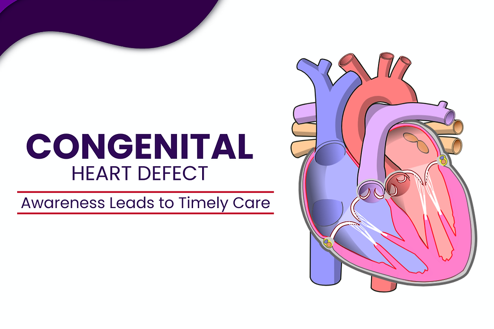
Ventricular Septal Defect (VSD) is one of the most frequently diagnosed forms of congenital heart defect (CHD). It refers to an opening in the wall (septum) that separates the heart’s two lower chambers, known as the ventricles. This structural difference is present from birth and affects how blood flows through the heart and lungs.
Among all congenital heart defects, VSD stands out not only because of its prevalence but also because of the wide range of ways it can present—from very small openings that close on their own to larger defects that require medical attention. Understanding VSD as part of the broader category of CHD helps explain why early recognition, careful evaluation, and appropriate monitoring are essential.
Understanding the Link Between VSD and Congenital Heart Disease (CHD)
Congenital heart disease includes any heart abnormality that develops before birth. VSD is classified under CHD because it forms during early fetal heart development, usually within the first few weeks of pregnancy when the heart chambers and walls are forming.
In a typical heart, oxygen-rich blood and oxygen-poor blood remain separated. A ventricular septal defect allows blood to pass from the left ventricle to the right ventricle, altering normal circulation. The impact of this depends on the size and location of the defect, as well as the pressure difference between the chambers.
Why Ventricular Septal Defect Occurs
The exact cause of VSD is not always identifiable. In most cases, it occurs when the ventricular septum does not form completely during fetal development. This can happen without any clear trigger.
However, certain factors are known to increase the likelihood of congenital heart defects, including VSD:
Genetic conditions or chromosomal variations
Family history of congenital heart disease
Maternal health conditions during pregnancy
Exposure to certain medications or infections in early pregnancy
It is important to note that VSD is not caused by anything done intentionally during pregnancy, and many cases occur in otherwise healthy pregnancies.
Types of Ventricular Septal Defect
VSD is not a single, uniform condition. It is categorized based on the location of the opening in the ventricular septum. Understanding these types helps explain why symptoms and management approaches can vary.
Perimembranous VSD: The most common type, located near the heart valves. Its position can influence valve function over time.
Muscular VSD: Found in the muscular portion of the septum. These defects are often small and may close naturally as the child grows.
Inlet VSD: Located near where blood enters the ventricles. This type is sometimes associated with other congenital heart conditions.
Outlet (Supracristal) VSD: Positioned near the exit of blood from the ventricles. Though less common, it may affect blood flow to major arteries.
Each type differs in clinical significance, which is why accurate diagnosis is essential.
Early Signs and Symptoms of VSD
The way VSD presents can vary significantly. Some individuals show signs early in infancy, while others may remain symptom-free for years, especially if the defect is small.
In infants, early indicators may include:
Difficulty feeding or tiring easily during feeds
Poor weight gain
Rapid or labored breathing
Frequent respiratory infections
In older children or adults, symptoms—if present—may be subtler and related to physical activity tolerance or fatigue. Small VSDs may produce no noticeable symptoms and are sometimes detected only during routine examinations.
How VSD Is Identified and Evaluated
The diagnostic process for ventricular septal defect focuses on understanding the structure of the heart and how blood is flowing through it. Evaluation usually begins when a healthcare provider detects a heart murmur during a physical examination.
Further assessment may involve:
Echocardiography: to visualize the heart’s structure and measure the size and location of the defect
Electrocardiogram (ECG): to assess heart rhythm and chamber workload
Chest imaging: to evaluate heart size and lung circulation
Advanced cardiac imaging: when needed, for detailed anatomical assessment
These tests help determine whether the defect is small or significant and guide decisions about monitoring or intervention.
How VSD Affects the Heart Over Time
The long-term impact of a ventricular septal defect depends on multiple factors, including defect size, location, and associated heart changes. Small VSDs often close spontaneously and may never affect heart function.
Larger defects can increase blood flow to the lungs, placing additional strain on the heart. If left unmonitored, this altered circulation may lead to complications over time. This is why regular follow-up is a key part of managing VSD as a congenital heart condition.
Management and Monitoring Approach
There is no single pathway for managing VSD. Many cases require only observation, especially when the defect is small and not causing symptoms. Others may benefit from medical management or corrective procedures based on clinical findings.
The goal is not only to address the defect but also to support normal growth, development, and long-term heart health. Decisions are always individualized and guided by qualified
cardiac specialists.
Why In-Depth, Reliable Information on VSD Matters
Ventricular septal defect is not uniform in how it appears or progresses. In some cases, the opening in the ventricular wall is small and allows the heart to function without noticeable strain. In others, the difference in blood flow leads to visible signs that develop over time rather than immediately at birth.
These variations explain why some individuals experience early symptoms while others remain unaffected for years. Diagnostic evaluation focuses not only on identifying the defect but also on understanding its size, location, and effect on circulation. When the heart adapts well, regular monitoring may be sufficient, while greater physiological impact requires closer
attention.
How the Condition Evolves
Ventricular septal defect represents a broad spectrum within congenital heart disease, ranging from minor findings to conditions requiring close attention. Its causes lie in early heart development, its types are anatomically distinct, and its effects depend on how the heart adapts over time.
A well-informed approach—grounded in accurate diagnosis, appropriate follow-up, and reliable education—remains central to managing VSD effectively. Understanding the condition in its entirety allows individuals and families to make informed decisions with confidence, guided by medical expertise rather than uncertainty.


Comments